Scientists at Scripps Research Center in La Jolla are working to streamline monoclonal antibody vaccine development by devising more efficient ways to screen for rare B antigen-specific B cells. [1]
Today, monoclonal antibodies (mAbs) are at the forefront of vaccine research as scientists continue to develop new treatments to fight cancer, autoimmunity, and infectious disease. Antigen-specific mAbs are particularly useful to vaccine science. These antibodies are designed to target surface proteins that are specific to a virus or pathological cell type, without harming healthy cells.
Of course, not all mAbs are equally efficient at fighting disease. Only a small percentage of the immune system’s mAb repertoire will have broad neutralizing capacity (these are known as broadly neutralizing antibodies or bnAbs). This is the capacity to recognize a wide range of viral variants, or even a whole family of related viruses, and to bind the target antigen with high affinity so that the invasive viral particle or cell can be destroyed efficiently.
Exceptionally potent antibodies like this make the most useful templates for vaccine development. However, they are also very rare, found in only a small subset of individuals exposed to a disease. This means that identifying high-value antibodies from a library of B cell antigens is both expensive and labor-intensive. The team at Scripps set their sights on closely examining current antibody screening techniques and determining which points could be optimized to develop a faster, more efficient approach.
Using DNA “barcoding”
The Scripps team understood that the most expensive, labor-intensive portion of mAb screening is the need to sequester single immortalized or transiently activated B cells from a large immune cell population. Until recently, this had to be accomplished manually through serial dilution. Luckily, recent advances in automation and microfluidics have made single-cell isolation less labor-intensive. There have also been advances in sequencing mAbs; CITE-seq [2] was developed as a method of performing RNA sequencing on surface proteins at the single-cell level. This technique pioneered the use of DNA “bar coding” through the use of oligonucleotide tags, so that each antibody has a unique molecular identifier, or UIM. LIBRA-seq [3], another tool in the mAb handbook, goes one step further by labeling single B cells (or T cells) before using them to generate a library of B cell receptor sequences that can be linked back to antigen specificity.
While incredibly useful, these techniques generate enormous amounts of data, with the result that analysis is still tedious and labor-intensive. The Scripps researchers’ mission, therefore, was to develop a next-generation LIBRA-seq methodology by optimizing B cell isolation and sorting while simultaneously developing an integrated computational framework for detailed B cell characterization.
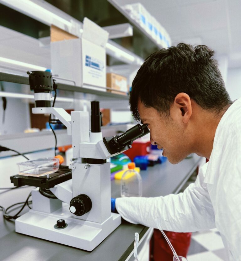
To enable their research, the scientists would first require high-quality cellular starting material. The peripheral blood mononuclear cells (PBMC) used for this study were isolated from highly characterized leukapheresis material collected from healthy donors at Excellos.
As part of their optimization studies, the researchers ran a series of experiments which made it clear that cell viability was a major factor affecting antigen-labeled B cell recovery. Bulk sorting of cells into 96-well plates containing aliquots of 100% FBS prior to library generation significantly improved labeled B cell viability and subsequent recovery efficiencies.
Enriching antigen-specific B cells
Antigen-barcode complexes (AgBCs) were next in line to be optimized. The original method for creating these complexes consisted of biotinylating protein antigens, conjugating them to barcode oligonucleotides, then using these pre-assembled complexes to label B cell receptors. Unfortunately, this meant that critical B cell epitopes are sometimes fully or partially blocked by the barcode oligonucleotide. To eliminate this drawback, the scientists added the barcoding reagent to site-specifically biotinylated protein antigens. This allowed each antigen to be linked to a barcoding reagent in a site-specific manner that minimizes binding interference with the native B cell receptor.
Selecting only B cells positive for both markers allowed for enhanced enrichment of antigen-specific B cells and a lower false positive background.
After optimizing their barcoding method, the researchers devised a dual antigen labeling strategy in which aliquots of the same antigens were labeled with two different fluorophore markers. Selecting only B cells positive for both markers allowed for enhanced enrichment of antigen-specific B cells and a lower false positive background.
Finally, the research team devised a computational analysis method integrating single B cell analysis and single-cell multi-omics data. This let the team streamline their output data and link binding affinity data to antigen-specific B cell receptors. This allowed for more efficient screening for highly potent bnAb. The researchers proceeded to demonstrate the utility of their work by using the new methodology to successfully identify ultra-rare HIV bnAb precursors.
This let the team streamline their output data and link binding affinity data to antigen-specific B cell receptors.
While the Scripps scientists would like to see further improvements made, they believe their work will streamline high-value mAb discovery and hence antibody-based vaccine development.
Excellos is proud to have supported this work. To learn about how we strive for excellence in our immunotherapy products and services, please contact us today.
Reference
- Jonathan Hurtado, Claudia Flynn, Jeong Hyun Lee, Eugenia Salcedo, Christopher A. Cottrell, Patrick D. Skog, David Nemazee, William R. Schief, Elise Landais, Devin Sok, Bryan Briney. Efficient isolation of rare B cells using next-generation antigen barcoding. bioRxiv 2022.06.06.495029; doi: https://doi.org/10.1101/2022.06.06.495029
- Stoeckius M, Hafemeister C, Stephenson W, Houck-Loomis B, Chattopadhyay PK,Swerdlow H, et al. Simultaneous epitope and transcriptome measurement in single cells. Nat Methods. 2017;14: 865–868.
- Setliff I, Shiakolas AR, Pilewski KA, Murji AA, Mapengo RE, Janowska K, et al. High-Throughput Mapping of B Cell Receptor Sequences to Antigen Specificity. Cell. 2019; 179:1636-1646.e15.